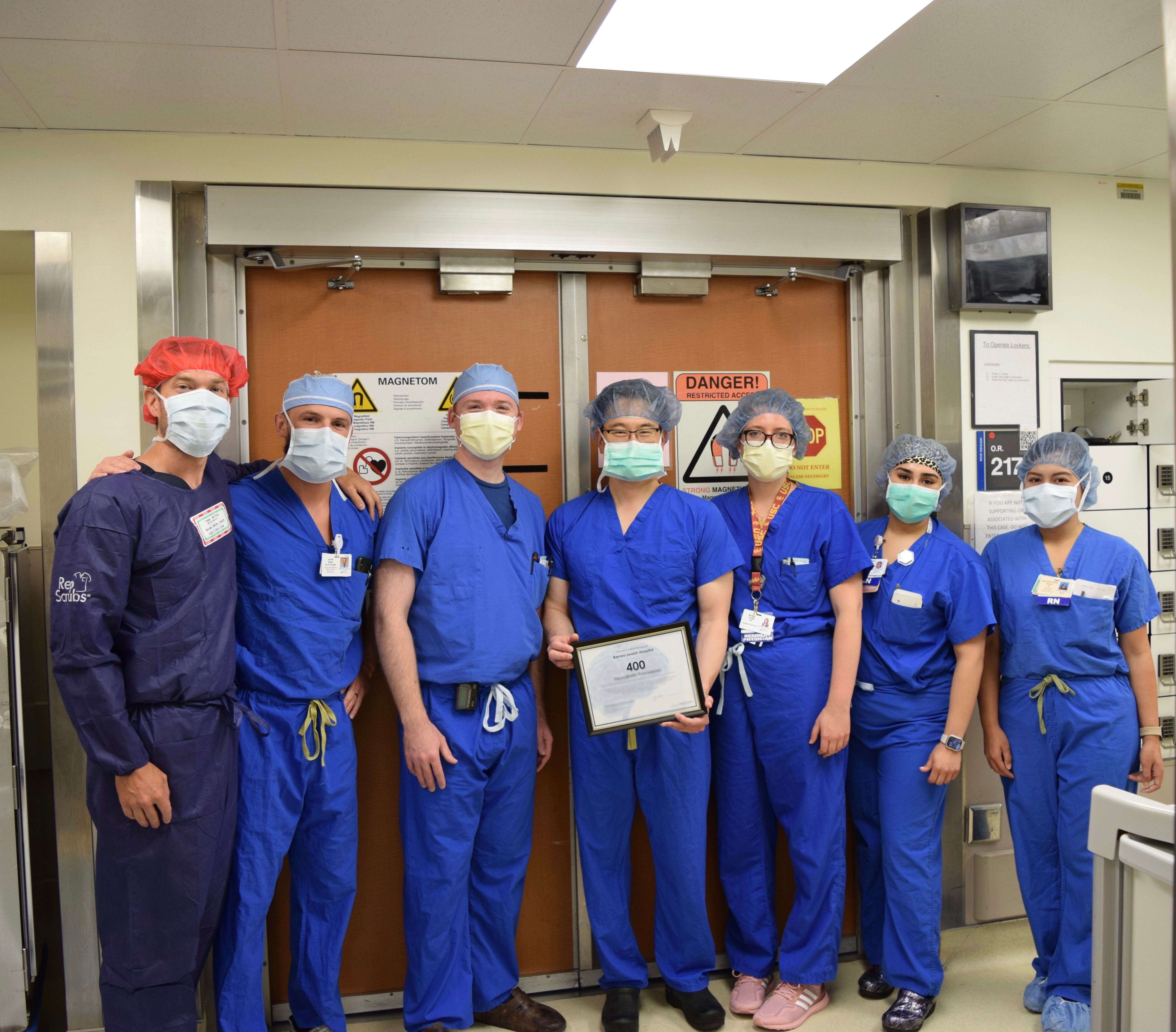
MRI-guided Neurosurgical Laser Ablation

Washington University neurosurgeons are among the first in the nation to use an MRI-guided, high-intensity laser probe designed especially for the treatment of “difficult to access” or surgically inoperable brain tumors.
At Washington University, use of this technology was pioneered by neurosurgeon Eric Leuthardt, MD, director of the Brain Laser Center and director of the Center for Innovation in Neuroscience and Technology.
The system utilizes a high-intensity laser probe to destroy cancer cells deep within the brain. Typically, tumors are in hard-to-reach regions of the brain such as the basal ganglia, thalamus and insula, although patients with cancer in other areas may also be considered for a number of clinical reasons.
The hallmark of the system is its ability to use magnetic resonance imaging (MRI) to precisely target laser-generated heat directly into a tumor to destroy cancer cells. The procedure leaves surrounding healthy brain tissue undamaged.
Several types of brain tumors can be treated with the Monteris system:
- Glial tumors, including gliomas
- Anaplastic astrocytomas
- Glioblastoma multiforme
- Metastatic cancers that have spread to the brain from other regions of the body
- Some radiation-resistant tumors
- Radiation necrosis due to prior radiation therapy
- Cause of epilepsy such as mesial temporal lobe epilepsy
How it works
Patients who are candidates for this procedure will have a small burr hole the diameter of a pencil drilled through their skull. Neurosurgeons then use real-time MR imaging to guide the probe through the brain and into the tumor. Once inside the tumor, the laser discharges highly focused thermal energy to coagulate and kill cancer cells.
Follow-up MRI studies of the tumor typically begin to show evidence of tumor death immediately after the surgery. The patient’s recovery usually takes several days but is generally much quicker than with open surgery.
Patients may not be eligible for treatment if either of the following applies:
- Patient is unable to get an MRI (e.g., patient has a pacemaker or other metal in their body).
- Patient’s tumor is located in an area that, if treated, could cause other significant neurological impairment (e.g., tumors in the brainstem).