Neuroendoscopy
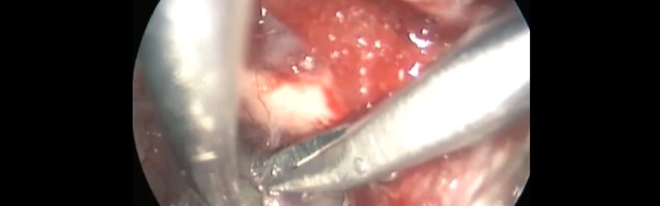
Neuroendoscopy involves the use of an endoscope—a long flexible tube that has a video camera and a light source—to perform diagnostic or therapeutic procedures in the brain, spine, or peripheral nervous system.
The endoscope can be equipped with specialized instruments to perform various procedures. Depending upon the location of a tumor(s), an endoscope can be maneuvered
- through the nose (for diseases of the skull base)
- through small incisions in the top of the head (for brain tumors or craniofacial conditions)
- through small incisions in the back (for spinal disease)
Endoscopic Endonasal Skull-Base Surgery
Many diseases of the skull base can be treated with endoscopic endonasal skull-base surgery. Traditionally, the surgical approach for these diseases was through facial incisions or through an opening of the scalp and then removal of a piece of the skull. In endoscopic endonasal skull-base surgery, however, the surgical team advances an endoscope through the nasal cavity to view the anatomy and perform the surgery. The approach eliminates the need to make incisions in the scalp and/or face.
Washington University neurosurgeons and otolaryngologists team with other specialists to perform endoscopic endonasal procedures, which can be for the treatment of
- Pituitary tumors
- Chordomas
- Chondrosarcomas
- Craniopharyngiomas
- Cysts
- Meningiomas
- Other types of brain tumors
- Cerebrospinal fluid leaks
Intraventricular Neuroendoscopy
- Colloid cysts – Neurosurgeons may use neuroendoscopy to remove colloid cysts, benign congenital tumors of the third ventricle that are rare and can cause headaches, vertigo, decreased memory, and behavioral changes.
- Biopsy – Neuroendoscopy is used for biopsy of tumors in the ventricles of the brain.
- Hydrocephalus – Endoscopic third ventriculostomies (ETVs) are used in the treatment of obstructive hydrocephalus. Obstructive hydrocephalus is a condition in which there is a build-up of cerebrospinal fluid (CSF) in the ventricles of the brain as a result of the blockage of CSF drainage. In ETV, the neurosurgeon makes a perforation in the floor of the third ventricle, allowing CSF to move out of the blocked ventricular system and into a neighboring CSF space. The goal of this procedure is to normalize pressure on the brain.